

Neurophysiology Essentials: A Complete Guide to Neuron Structure, Synapses, and Nervous System Functions
1.What is neuron? Explain the functions of dendrite and axon terminals of a neuron.
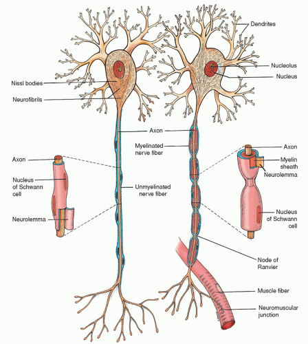
A neuron is the fundamental signaling cell of the nervous system, specialized both to receive incoming information, integrate it, and transmit signals onward. Structurally, most neurons have three principal parts:
Cell body (soma) – contains the nucleus, Nissl bodies (rough ER), ribosomes, mitochondria and the cytoskeleton (neurofibrils and microtubules).
Dendrites – multiple, branched “little trees” that extend from the soma.
Axon – a single long process arising from the axon hillock that conducts action potentials to its terminals.
Cell Body (Soma):
Contains the nucleus, rough ER (Nissl bodies), ribosomes, mitochondria, and a cytoskeleton of neurofibrils and microtubules.
Makes proteins needed for cell function, growth, and repair.
Gathers incoming signals but does not fire action potentials itself.
Dendrites:
Many short, tree-like branches off the cell body, often with tiny “spines.”
Have receptors (ligand-gated channels and G‑protein receptors) that bind neurotransmitters.
When channels open, they produce small local voltage changes (EPSPs or IPSPs) that travel toward the cell body.
All these inputs add up at the axon hillock; if the sum reaches about –55 mV, an action potential starts.
Axon and Axon Terminals:
A single long fiber (axon) carries a rapid, all‑or‑nothing electrical impulse away from the cell body.
The axon splits into many endings (axon terminals or telodendria), each with vesicles of neurotransmitter and SNARE proteins.
When the impulse arrives, voltage‑gated Ca²⁺ channels open, Ca²⁺ enters, and vesicles release their neurotransmitter into the synapse.
Afterward, neurotransmitters are cleared by enzymes or taken back up into the terminal for reuse.
2. Illustrate the structure and functions of an axon in a neuron.
Structure and Functions of an Axon
Structure of an Axon:
The axon is a long, thin extension of the neuron.
It starts at the axon hillock (a cone-shaped area of the cell body).
Inside, it contains axoplasm (the cytoplasm), mitochondria, and microtubules (which help transport materials).
It is covered by a membrane called the axolemma.
The axon may have branches called axon collaterals.
It ends in axon terminals, which have synaptic end bulbs containing neurotransmitters.
Functions of an Axon:
Conducts Nerve Impulses:
Carries electrical signals (action potentials) from the cell body to other neurons, muscles, or glands.Triggers Neurotransmitter Release:
When the signal reaches the axon terminal, it causes the release of chemicals (neurotransmitters) into the synapse.Transports Materials:
Moves nutrients, proteins, and waste between the cell body and the axon terminals using fast and slow axonal transport.Communication:
Helps the neuron send messages to other cells across synapses.
3.Describe the structural and functional classification of neurons.
Structural and Functional Classification of Neurons
1. Structural Classification
This is based on the number of processes (branches) coming out of the cell body.
a) Multipolar Neurons
Structure: One axon, many dendrites.
Example: Most neurons in the brain and spinal cord.
Function: Common in motor and interneurons.
b) Bipolar Neurons
Structure: One axon, one dendrite.
Example: Found in the retina (eye), inner ear, and nose.
Function: Sensory neurons for special senses.
c) Unipolar (Pseudounipolar) Neurons
Structure: One branch that splits into two – one goes to the body, the other to the spinal cord.
Example: Found in sensory neurons of the skin, joints, and muscles.
Function: Carries touch, pain, and temperature signals to the CNS.
2. Functional Classification
a) Sensory Neurons (Afferent)
Function: Carry signals from the body (like skin, eyes) to the brain and spinal cord.
Example: Touch or pain sensors.
b) Motor Neurons (Efferent)
Function: Carry signals from the brain and spinal cord to muscles or glands.
Example: Cause your hand to move.
c) Interneurons (Association Neurons)
Function: Connect sensory and motor neurons inside the brain or spinal cord.
Example: Involved in reflexes and decision-making.
4.What are the astrocytes? Illustrate how do astrocytes play the following role in CNS:
a) Potassium buffer
b) Removing excess neurotransmitters
c) Glycogen reserve
Astrocytes are star-shaped glial (support) cells found in the central nervous system (CNS) — the brain and spinal cord. They help neurons work properly and keep the brain environment balanced.
Roles of Astrocytes in the CNS:
a) Potassium Buffer
During nerve activity, neurons release potassium ions (K⁺) into the space around them.
Too much K⁺ can be harmful.
Astrocytes absorb the extra K⁺ to keep the balance of ions and prevent overexcitation of neurons.
b) Removing Excess Neurotransmitters
After a neuron sends a message, neurotransmitters like glutamate remain in the synapse.
Astrocytes take in these extra neurotransmitters to stop the signal and protect neurons from damage.
c) Glycogen Reserve
Astrocytes store glycogen, a form of energy.
When neurons need extra energy (like during high activity or low glucose), astrocytes break down glycogen and provide glucose to support the neurons.
5. Describe the structure and function of blood-brain barrier and blood-CSF barrier.
Blood-Brain Barrier (BBB)
Structure:
Made of tight junctions between capillary walls in the brain.
Supported by astrocytes (a type of glial cell) that help maintain the barrier.
The walls are made of endothelial cells that are tightly packed, so very few things can pass through.
Function:
Protects the brain by blocking harmful substances (like toxins, bacteria) in the blood from entering brain tissue.
Allows only certain needed substances (like oxygen, glucose) to pass.
Maintains a stable environment for brain function.
Blood-CSF Barrier
Structure:
Found in the choroid plexus (where cerebrospinal fluid is made).
Made of ependymal cells connected by tight junctions.
Also includes capillaries that filter blood to form CSF.
Function:
Controls what enters the cerebrospinal fluid (CSF) from the blood.
Keeps out harmful substances while allowing useful ones like nutrients to pass.
Helps maintain the chemical balance of CSF around the brain and spinal cord.
6. Define myelination. Describe the mechanism of myelination by Schwann cells in PNS
Myelination is the process where certain glial cells wrap around the axons of neurons with a fatty layer called myelin. This myelin acts like insulation, helping nerve signals travel faster and more efficiently.
Myelination by Schwann Cells in the PNS (Peripheral Nervous System)
Mechanism :
A Schwann cell attaches to a part of a neuron’s axon.
The Schwann cell starts to wrap its membrane around the axon many times, like rolling tape.
Each wrap adds more layers of myelin, forming a thick insulating covering.
The inside part of the Schwann cell becomes the myelin sheath, and the outermost part (with the nucleus) is called the neurilemma.
Gaps between Schwann cells along the axon are called Nodes of Ranvier — these help nerve signals jump quickly from one gap to the next (called saltatory conduction).
7. Define synapse. Describe the structure, locations and the advantages of an electrical synapse.
A synapse is the place where a nerve cell (neuron) connects with another neuron, muscle, or gland cell to pass messages. It’s like a small gap where signals are shared.
Types of Synapses:
There are two main types of synapses:
Chemical Synapse – Uses chemicals (neurotransmitters) to send signals.
Electrical Synapse – Sends signals directly using electric current.
Structure of an Electrical Synapse:
Very small gap between two neurons (only about 3.5 nanometers).
Neurons are connected by gap junctions (like tiny tunnels).
These junctions allow ions (charged particles) to pass directly from one cell to another.
No neurotransmitters are needed.
Location of Electrical Synapses:
Found in some areas of the brain.
Found in the heart and some smooth muscles (like intestines).
Also seen in developing nervous systems (in embryos).
Advantages of Electrical Synapses:
Very fast — signal passes instantly.
Two-way communication — signals can go both directions.
Good for actions that need to happen quickly and together (like heartbeats or reflexes).
Helps groups of neurons work synchronously (at the same time).
8.What is a chemical synapse? Illustrate the mechanism of signal transmission through a typical chemical synapse.
A chemical synapse is a junction between two neurons where signals are passed using chemicals called neurotransmitters. Unlike electrical synapses, which use direct electrical connections, chemical synapses need chemicals to carry the signal across the gap (synaptic cleft).
Mechanism of Signal Transmission Through a Chemical Synapse:
Arrival of Action Potential:
When a nerve signal (action potential) reaches the axon terminal of a neuron, it triggers the next steps.Calcium Influx:
The action potential causes voltage-gated calcium channels to open. Calcium ions (Ca²⁺) flow into the axon terminal.Neurotransmitter Release:
The calcium ions cause synaptic vesicles (tiny sacs inside the axon terminal) to move and fuse with the cell membrane. This releases the neurotransmitters into the synaptic cleft (the gap between the two cells).Binding to Receptors:
The neurotransmitters travel across the synaptic cleft and bind to specific receptors on the postsynaptic cell (the next neuron, muscle, or gland cell).Effect on Postsynaptic Cell:
When neurotransmitters bind to the receptors, they can either:Open ion channels, leading to changes in the membrane potential (excitation or inhibition).
Cause a new action potential if the signal is strong enough.
Neurotransmitter Removal:
After the signal is sent, the neurotransmitters need to be removed from the synapse. This can happen by:Reuptake: The neurotransmitter is taken back into the presynaptic cell to be reused.
Degradation: An enzyme breaks down the neurotransmitter in the synaptic cleft.
9. Describe the structure and function of an electrical synapse. "The electrical synapses are faster than chemical synapses"- why.
An electrical synapse is a connection between two neurons where the signal is passed directly by electric current. It is different from a chemical synapse because it doesn’t need neurotransmitters to transmit signals.
Structure of an Electrical Synapse:
Very small gap between two neurons (only about 3.5 nanometers).
Neurons are connected by gap junctions (like tiny tunnels).
These junctions allow ions (charged particles) to pass directly from one cell to another.
No neurotransmitters are needed.
Function of an Electrical Synapse:
Fast Signal Transmission:
The electric signal travels quickly between cells, allowing the neurons to communicate almost instantly.Two-Way Communication:
Unlike chemical synapses, the signal in electrical synapses can travel in both directions, allowing fast feedback.Synchronized Activity:
Electrical synapses help groups of neurons work together in a coordinated manner, like in the heart where all muscle cells need to contract at the same time.
Here are the reasons why Electrical Synapses Faster Than Chemical Synapses.
No Delay for Chemical Release:
In chemical synapses, the signal has to wait for neurotransmitters to be released, travel across the synapse, and bind to receptors. This takes time.
In electrical synapses, the electric current flows directly through the gap junctions, so there’s no delay. The signal is almost instantaneous.


0 Comments